During their summer research, Kaitlyn Robles-Patterson ’27 and Dr. Smith used advanced imaging to reconstruct tissue structures inside the inner ear.
By
Sara Colabella ’08, MA’11, EdD’26
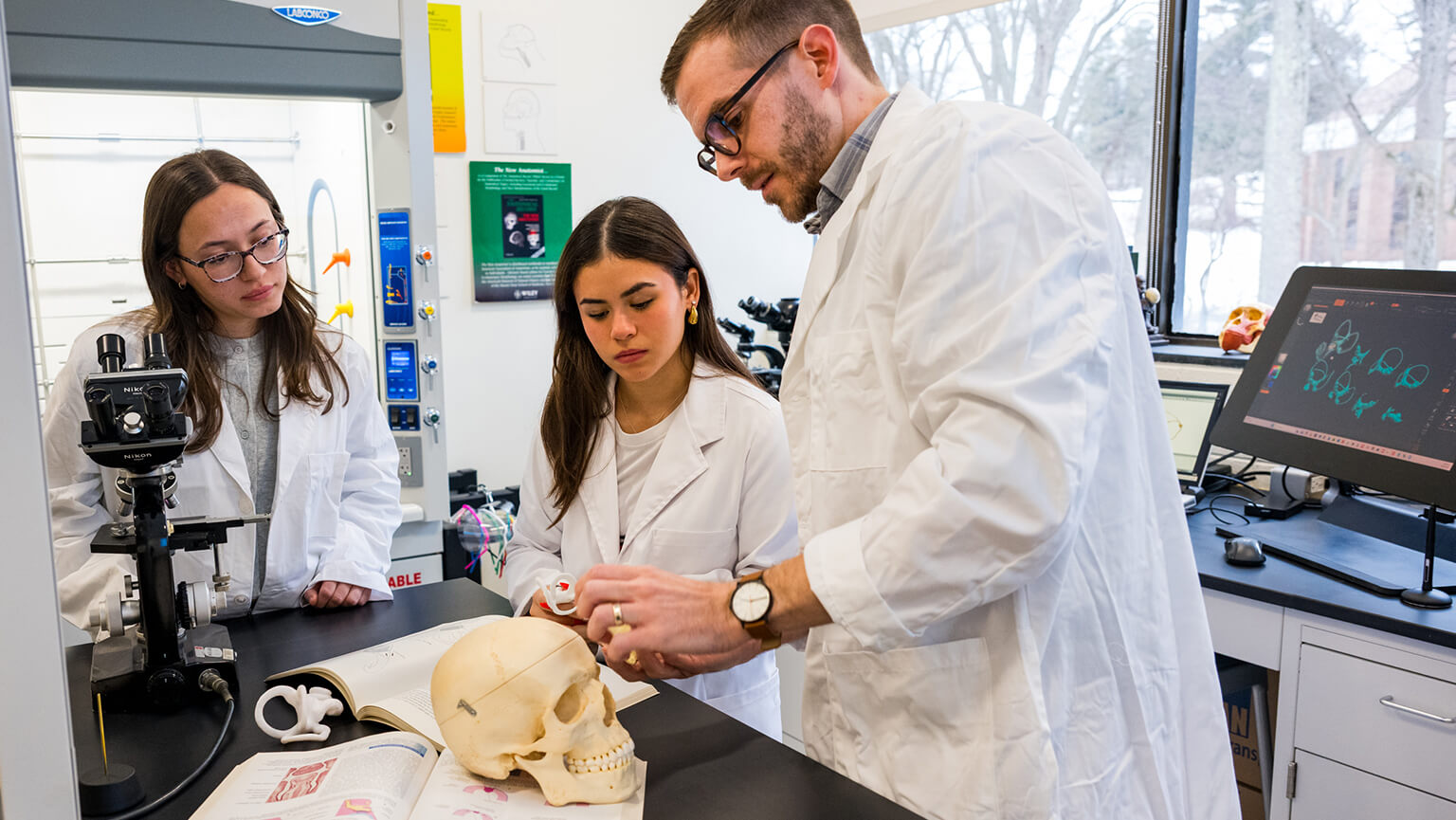
Student researchers in Dr. Christopher Smith’s lab use CT scans to model the inner ear of a gibbon.
Using advanced imaging and the skills of an artist, a faculty-student research team brings the hidden structures of the inner ear into view.
Deep within the human skull, tucked inside a space the size of a Tic Tac, is a complex system that allows us to hear, maintain our balance, and navigate the world around us. Despite centuries of study and advances in medical imaging, scientists know little about the delicate structures hidden within the inner ear.
Christopher Smith, PhD, assistant professor of biology at the John Charles Meditz College of Arts and Sciences, is working to change that. An evolutionary anatomist with a background in medical illustration, Dr. Smith uses advanced imaging techniques, three-dimensional modeling, and comparative anatomy to investigate how hearing and balance systems evolved, how they function, and how subtle variations in their anatomy may contribute to disorders such as vertigo and hearing loss.
“It’s an incredible area of the body,” Dr. Smith said. “These structures are central to how we experience the world, how we orient ourselves in space, how we move, and how we hear.”
We know a tremendous amount about the cellular and genetic levels of the ear, but there is still a black box when it comes to understanding the overall anatomy of these membranous structures."
– Christopher Smith, PhD, Assistant Professor of Biology
Over the summer, undergraduate researcher Kaitlyn Robles-Patterson ’27 assisted Dr. Smith in reconstructing previously unseen anatomical structures within the inner ear. “We looked at the inner ear anatomy of primates and compared it to human anatomy,” said Robles-Patterson, “to better understand how anatomy may relate to locomotion, sensitivity, and behavior.”
The project focused on gibbons, a group of small apes known for their distinctive vocalizations and remarkable agility in the forest canopy. “One of the things that Katie’s project did was to dig into these gibbon scans that we have, reconstructing not only more of the bony morphology, but the membranes inside that can help us better understand the anatomy,” said Dr. Smith.
Dr. Smith’s path to research began in the art studio. As an undergraduate studying exercise science, he initially envisioned a career in athletic training or physical therapy. But he also had a lifelong passion for drawing and painting. Looking for a way to unite his interests in art and anatomy, he enrolled in the medical and biological illustration master’s program at Johns Hopkins University.
“Art was the gateway into science for me,” Dr. Smith said. “If it wasn’t for my experience in art, I probably would not have pursued research.”
Today, his artistic training remains central to his work. Many of the structures Dr. Smith studies are too small to visualize fully even using the most advanced medical imaging technologies. Researchers can readily image bone, but many of the soft tissues within the inner ear remain elusive.
Student researchers in Dr. Christopher Smith’s lab use CT scans to model the inner ear of a gibbon.
Jaclyn Cusa ’27 at the microscope.
“We know a tremendous amount about the cellular and genetic levels of the ear,” Dr. Smith explained, “but there is still a black box when it comes to understanding the overall anatomy of these membranous structures.”
Drawing on the observational and visualization skills he developed as an artist, Dr. Smith creates detailed three-dimensional reconstructions using high-resolution CT scans and digital modeling techniques. These visualizations reveal anatomy that would otherwise be inaccessible, enabling researchers to ask new questions about how inner ear structures function and how they evolved.
One area of his research examines whether variations in inner ear anatomy contribute to disorders such as vertigo and Meniere’s disease. Though millions of people experience balance-related disorders, the anatomical factors behind those conditions are not well-known.
“There are certain structures that we simply have not been able to visualize well enough to fully understand their role in disease,” Dr. Smith said.
Another area of Dr. Smith’s work examines how sensory systems have evolved throughout human and primate history. By studying both living species and fossil remains, he hopes to shed light on how hearing and balance systems developed alongside larger brains, altered skull shapes, and the evolution of upright walking.
His research is helping address a longstanding challenge in evolutionary biology: understanding soft tissues that no longer exist. Currently, fossil-related studies rely on preserved bone. Dr. Smith and his collaborators are developing methods to reconstruct the soft tissue anatomy to provide a more complete picture of how extinct species experienced their environments.
“All we usually have is the hard tissue,” said Dr. Smith. “The challenge is figuring out what the soft tissues looked like and how they functioned.”
To enhance his research with humans and primates, he is also building a collection of whale specimens to study how hearing and balance systems evolved in aquatic mammals. “Many whale species are highly intelligent, highly social mammals, similar to humans, but they evolved in completely different environments,” he said. “That makes them fascinating comparative models.” For Dr. Smith, mentoring students is one of the most rewarding aspects of his work. His lab is centered on student research with four to five students working alongside him each semester. “When I was an undergraduate, I had no idea research like this existed,” he said. “Being able to expose students to these opportunities and show them different paths into science is really exciting.”